Table of contents
Volume 5, Issue 7, pp. 89 - 118, July 2021
Cover: This month in
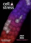
Cell Stress: The neuro-developmental role of autophagy. Image depicts mosaic expression of RNAi constructs and GFP reporter (light cells) in Drosophila fat body, a tissue, which shows programmed autophagy during development. Fluorescent autophagy reporter constructs (red-purple) are used to compare the autophagy phenotype of RNAi-expressing cells to that of surrounding cells. Image Credit: Andreas Zimmermann. The cover is published under the
CC BY 4.0 license.
Enlarge issue cover
Elevated plasma levels of the appetite-stimulator ACBP/DBI in fasting and obese subjects
Sijing Li, Adrien Joseph, Isabelle Martins and Guido Kroemer
Viewpoint |
page 89-98 | 10.15698/cst2021.07.252 | Full text | PDF |
Abstract
Eukaryotic cells release the phylogenetically ancient protein acyl coenzyme A binding protein (ACBP, which in humans is encoded by the gene DBI, diazepam binding inhibitor) upon nutrient deprivation. Accordingly, mice that are starved for one to two days and humans that undergo voluntary fasting for one to three weeks manifest an increase in the plasma concentration of ACBP/DBI. Paradoxically, ACBP/DBI levels also increase in obese mice and humans. Since ACBP/DBI stimulates appetite, this latter finding may explain why obesity constitutes a self-perpetuating state. Here, we present a theoretical framework to embed these findings in the mechanisms of weight control, as well as a bioinformatics analysis showing that, irrespective of the human cell or tissue type, one single isoform of ACBP/DBI (ACBP1) is preponderant (~90% of all DBI transcripts, with the sole exception of the testis, where it is ~70%). Based on our knowledge, we conclude that ACBP1 is subjected to a biphasic transcriptional and post-transcriptional regulation, explaining why obesity and fasting both are associated with increased circulating ACBP1 protein levels.
Towards a better understanding of the neuro-developmental role of autophagy in sickness and in health
Juan Zapata-Muñoz, Beatriz Villarejo-Zori, Pablo Largo-Barrientos and Patricia Boya
Reviews |
page 99-118 | 10.15698/cst2021.07.253 | Full text | PDF |
Abstract
Autophagy is a critical cellular process by which biomolecules and cellular organelles are degraded in an orderly manner inside lysosomes. This process is particularly important in neurons: these post-mitotic cells cannot divide or be easily replaced and are therefore especially sensitive to the accumulation of toxic proteins and damaged organelles. Dysregulation of neuronal autophagy is well documented in a range of neurodegenerative diseases. However, growing evidence indicates that autophagy also critically contributes to neurodevelopmental cellular processes, including neurogenesis, maintenance of neural stem cell homeostasis, differentiation, metabolic reprogramming, and synaptic remodelling. These findings implicate autophagy in neurodevelopmental disorders. In this review we discuss the current understanding of the role of autophagy in neurodevelopment and neurodevelopmental disorders, as well as currently available tools and techniques that can be used to further investigate this association.


