Table of contents
Volume 5, Issue 2, pp. 23 - 28, February 2021
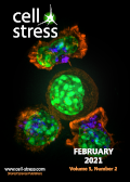
Cover: This month in
Cell Stress: RIG-I signaling against cancer T-cell resistance. Fluorescence microscopy image of killer T-cells attacking a cancer cell (center). Credit: Alex Ritter, Jennifer Lippincott Schwartz and Gillian Griffiths, National Institutes of Health. Public domain image modified by
Cell Stress. The cover is published under the
CC BY 4.0 license.
Enlarge issue cover
Innate RIG-I signaling restores antigen presentation in tumors and overcomes T cell resistance
Beatrice Thier and Annette Paschen
Microreviews |
page 26-28 | 10.15698/cst2021.02.242 | Full text | PDF |
Abstract
In recent years, therapy with immune modulating antibodies, termed immune checkpoint blockade (ICB), has revolutionized the treatment of advanced metastatic melanoma, yielding long-lasting clinical responses in a subgroup of patients. But despite this remarkable progress, resistance to therapy represents a major clinical challenge. ICB efficacy is critically dependent on cytotoxic CD8+ T cells targeting tumor cells in an HLA class I (HLA-I) antigen-dependent manner. Transcriptional suppression of the HLA-I antigen processing and presentation machinery (HLA-I APM) in melanoma cells leads to HLA-I-low/-negative tumor cell phenotypes escaping CD8+ T cell recognition and contributing to ICB resistance. In general, HLA-I-low/-negative tumor cells can be re-sensitized to T cells by interferons (IFN), augmenting HLA-I APM expression. However, this mechanism fails when melanoma cells acquire resistance to IFN, which recently turned out as a key resistance mechanism in ICB, besides HLA-I APM suppression. Seeking for a strategy to overcome these barriers, we identified a novel mechanism that restores HLA-I antigen presentation in tumor cells independent of IFN (Such et al. (2020) J Clin Invest, doi: 10.1172/JCI131572). We demonstrated that tumor cell-intrinsic activation of the cytosolic innate immunoreceptor RIG-I by its synthetic ligand 3pRNA overcomes transcriptional HLA-I APM suppression in patient-derived IFN-resistant melanoma cells. De novo HLA-I APM expression is IRF1/IRF3-dependent and re-sensitizes melanoma cells to autologous cytotoxic CD8+ T cells. Notably, synthetic RIG-I ligands and ICB synergize in T cell activation, suggesting combinational therapy could be an efficient strategy to improve patient outcomes in melanoma.
Mitochondrial dynamics links PINCH-1 signaling to proline metabolic reprogramming and tumor growth
Ling Guo and Chuanyue Wu
Microreviews |
page 23-25 | 10.15698/cst2021.02.241 | Full text | PDF |
Abstract
Proline metabolism is critical for cellular response to microenvironmental stress in living organisms across different kingdoms, ranging from bacteria, plants to animals. In bacteria and plants, proline is known to accrue in response to osmotic and other stresses. In higher organisms such as human, proline metabolism plays important roles in physiology as well as pathological processes including cancer. The importance of proline metabolism in physiology and diseases lies in the fact that the products of proline metabolism are intimately involved in essential cellular processes including protein synthesis, energy production and redox signaling. A surge of protein synthesis in fast proliferating cancer cells, for example, results in markedly increased demand for proline. Proline synthesis is frequently unable to meet the demand in fast proliferating cancer cells. The inadequacy of proline or “proline vulnerability” in cancer may provide an opportunity for therapeutic control of cancer progression. To this end, it is important to understand the signaling mechanism through which proline synthesis is regulated. In a recent study (Guo et al., Nat Commun 11(1):4913, doi: 10.1038/s41467-020-18753-6), we have identified PINCH-1, a component of cell-extracellular matrix (ECM) adhesions, as an important regulator of proline synthesis and cancer progression.


