Table of contents
Volume 3, Issue 1, pp. 1 - 28, January 2019
Cover: This month in
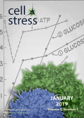
Cell Stress: Metabolic stress in antitumor immune response. Scanning electron micrograph of human T lymphocytes from the immune system of a healthy donor. Credit: NIAID. Background image from page 239 of "Ciba Foundation Symposium on the Regulation of Cell Metabolism" (1959, public domain). Image modified by
Cell Stress. The cover is published under the
CC BY 4.0 license.
Enlarge issue cover
Control of nucleolar stress and translational reprogramming by lncRNAs
Yvessa Verheyden, Lucas Goedert and Eleonora Leucci
Reviews |
page 19-26 | 10.15698/cst2019.01.172 | Full text | PDF |
Abstract
Under adverse environmental conditions, cells activate stress responses that favour adaptation or, in case of irreversible damage, induce cell death. Multiple stress response pathways converge to downregulate ribosome biogenesis and translation since these are the most energy consuming processes in the cell. This adaptive response allows preserving genomic stability and saving energy for the recovery. It follows that the nucleolus is a major sensor and integrator of stress responses that are then transmitted to the translation machinery through an intricate series of conserved events. Long non-coding RNAs (lncRNAs) are emerging as important regulators of stress-induced cascades, for their ability to mediate post-transcriptional responses. Consistently, many of them are specifically expressed under stress conditions and a few have been already functionally linked to these processes, thus further supporting a role in stress management. In this review we survey different archetypes of lncRNAs specifically implicated in the regulation of nucleolar functions and translation reprogramming during stress responses.
Regulation of T cell antitumor immune response by tumor induced metabolic stress
Fanny Chalmin, Mélanie Bruchard, Frederique Vegran and Francois Ghiringhelli
Reviews |
page 9-18 | 10.15698/cst2019.01.171 | Full text | PDF |
Abstract
Adaptive T cell immune response is essential for tumor growth control. The efficacy of immune checkpoint inhibitors is regulated by intratumoral immune response. The tumor microenvironment has a major role in adaptive immune response tuning. Tumor cells generate a particular metabolic environment in comparison to other tissues. Tumors are characterized by glycolysis, hypoxia, acidosis, amino acid depletion and fatty acid metabolism modification. Such metabolic changes promote tumor growth, impair immune response and lead to resistance to therapies. This review will detail how these modifications strongly affect CD8 and CD4 T cell functions and impact immunotherapy efficacy.
Serum- and glucocorticoid-inducible kinase 1 and the response to cell stress
Florian Lang, Christos Stournaras, Nefeli Zacharopoulou, Jakob Voelkl, Ioana Alesutan
Reviews |
page 1-8 | 10.15698/cst2019.01.170 | Full text | PDF |
Abstract
Expression of the serum- and glucocorticoid-inducible kinase 1 (SGK1) is up-regulated by several types of cell stress, such as ischemia, radiation and hyperosmotic shock. The SGK1 protein is activated by a signaling cascade involving phosphatidylinositide-3-kinase (PI3K), 3-phosphoinositide-dependent kinase 1 (PDK1) and mammalian target of rapamycin (mTOR). SGK1 up-regulates Na+/K+-ATPase, a variety of carriers including Na+-,K+-,2Cl–– cotransporter (NKCC), NaCl cotransporter (NCC), Na+/H+ exchangers, diverse amino acid transporters and several glucose carriers such as Na+-coupled glucose transporter SGLT1. SGK1 further up-regulates a large number of ion channels including epithelial Na+ channel ENaC, voltage-gated Na+ channel SCN5A, Ca2+ release-activated Ca2+ channel (ORAI1) with its stimulator STIM1, epithelial Ca2+ channels TRPV5 and TRPV6 and diverse K+ channels. Furthermore, SGK1 influences transcription factors such as nuclear factor kappa-B (NF-κB), p53 tumor suppressor protein, cAMP responsive element-binding protein (CREB), activator protein-1 (AP-1) and forkhead box O3 protein (FOXO3a). Thus, SGK1 supports cellular glucose uptake and glycolysis, angiogenesis, cell survival, cell migration, and wound healing. Presumably as last line of defense against tissue injury, SGK1 fosters tissue fibrosis and tissue calcification replacing energy consuming cells.
P4HA1 is a new regulator of the HIF-1 pathway in breast cancer
Ren Xu
Microreviews |
page 27-28 | 10.15698/cst2019.01.173 | Full text | PDF |
Abstract
Hypoxia-Inducible Factor (HIF)-1 is a transcription factor that plays the key role in response to low oxygen concentrations, or hypoxia. Activation of the HIF-1 pathway is not only crucial for normal tissue development and function, but also involved in disease progression, such as cancer. Cancer cells proliferate rapidly in solid tumors, and thus, solid tumors consume much more oxygen and nutrients than normal tissue and generate oxygen tension. It is well established that oxygen tension in solid tumor tissue induces the aberrant activation of the HIF-1 pathway, and subsequently promotes angiogenesis and tumor progression. Breast cancer is a heterogeneous disease and can be classified into luminal, Her2 positive, and triple negative (TNBC) subtypes base on ER/PR and Her2 expression in the breast cancer tissue. HIF-1 activation is observed in all breast cancer subtypes; interestingly, the HIF-1 pathway is hyperactivated in TNBCs compared to other subtypes. The differential activation of the HIF-1 pathway in breast cancer subtypes suggests that oxygen-independent pathways may be involved in HIF-1 regulation during TNBC progression. However, these pathways have not been well-characterized. We demonstrate that collagen proly 4 hydroxylase 1 (P4H1) induces HIF-1α expression at the protein level by modulating α-ketoglutarate (α-KG) and succinate levels. These results reveal a novel link between collagen hydroxylation and activation of the HIF-1 pathway and unveil a new HIF-1 regulation mechanism in TNBC.


