Table of contents
Volume 2, Issue 5, pp. 96 - 121, May 2018
Cover: This month in
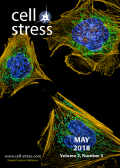
Cell Stress: Rad51-dependent DNA repair and genome stability. Image depicts fibroblasts with nuclei in blue, mitochondria in green, and the actin cytoskeleton in dark yellow. Credit: D. Burnette, J. Lippincott-Schwartz/NICHD; licensed under the
CC BY 2.0 license. Image modified by
Cell Stress. The cover is published under the
CC BY 4.0 license.
Enlarge issue cover
Putting together and taking apart: assembly and disassembly of the Rad51 nucleoprotein filament in DNA repair and genome stability
Tadas Andriuskevicius, Oleksii Kotenko and Svetlana Makovets
Reviews |
page 96-112 | 10.15698/cst2018.05.134 | Full text | PDF |
Abstract
Homologous recombination is a key mechanism providing both genome stability and genetic diversity in all living organisms. Recombinases play a central role in this pathway: multiple protein subunits of Rad51 or its orthologues bind single-stranded DNA to form a nucleoprotein filament which is essential for initiating recombination events. Multiple factors are involved in the regulation of this step, both positively and negatively. In this review, we discuss Rad51 nucleoprotein assembly and disassembly, how it is regulated and what functional significance it has in genome maintenance.
Calling RNF168 to action
Somaira Nowsheen and Zhenkun Lou
Microreviews |
page 113-114 | 10.15698/cst2018.05.135 | Full text | PDF |
Abstract
Genomic stress leads to various forms of DNA damage, of which DNA double strand breaks (DSBs) are the most lethal. An army of signaling molecules is called to action as soon as these DNA breaks are detected. Various protein modifications, such as phosphorylation and ubiquitination, are an integral part of the reaction. While phosphorylation activates various proteins, ubiquitin (Ub) adducts typically act as docking sites for DNA repair factors. The response to DNA DSB starts with the protein kinase ATM phosphorylating various substrates including MDC1 and histone H2AX. This mediator protein, MDC1, then recognizes phosphorylated histone H2AX and amplifies the damage response. The E3 ligase, RNF8, recognizes and binds to phosphorylated MDC1. RNF8 then modifies an unknown protein to call a second ubiquitin ligase, RNF168, into action. It has been recognized that these two ubiquitin ligases are recruited sequentially but there is an unknown linker protein between them. These two ubiquitin ligases are crucial to the formation of DSB-associated ubiquitin conjugates and, as a result, there has been long standing interest in the field in identifying the link between the two factors. In this paper we identify lethal(3) malignant brain tumor like 2 (L3MBTL2) as the substrate of RNF8 (Nowsheen S, et al. Nat Cell Biol 20:455-464, 2018). We report that ATM-mediated phosphorylation of the polycomb group like protein L3MBTL2 and subsequent interaction with MDC1 brings it to the vicinity of the DNA lesion. RNF8 acts upon this phosphorylated L3MBTL2 and generates K63-linked polyubiquitin chains. This modified substrate is subsequently recognized by RNF168 and tethers the protein to the DNA lesion. RNF168 then ubiquitinates proteins such as histone H2A and H2AX to further amplify the damage response and recruit repair proteins such as BRCA1 and 53BP1 (Figure 1).
Endolysosomal dysfunction and exosome secretion: implications for neurodegenerative disorders
André M. Miranda and Gilbert Di Paolo
Microreviews |
page 115-118 | 10.15698/cst2018.05.136 | Full text | PDF |
Abstract
Growing evidence suggests that endolysosomal and autophagic defects are key pathogenic processes in various neurodegenerative disorders, including Alzheimer’s disease (AD), Parkinson’s disease (PD), frontotemporal dementia (FTD) and amyotrophic lateral sclerosis (ALS). The causal relationship between these defects and neurodegeneration is supported by human genetic studies identifying disease mutations in genes controlling endolysosomal function and autophagy. The canonical view is that defects in these processes lead to impaired lysosomal clearance of proteins prone to form toxic oligomeric assemblies and/or aggregates, ultimately resulting in cellular pathologies that define these disorders. Because lysosomes mediate the clearance of a large number of lipids, lipid storage is frequently associated with compromised endolysosomal and autophagic function. However, an emerging notion, supported by our recent study on class III phosphatidylinositol 3-kinase (PI3K) Vps34, is that neuronal endolysosomal and autophagic dysfunction can manifest itself with the occurrence of physically damaged endomembranes and with the release of exosomes enriched for Amyloid Precursor Protein COOH-terminal fragments (APP-CTFs) as well as atypical phospholipid bis(monoacylglycero)phosphate (BMP). Here, we summarize our recent findings and their potential implications in the context of lysosomal biology, lipid signaling and neurodegenerative diseases.
Exosomal secretion of truncated cytosolic lysyl-tRNA synthetase induces inflammation during cell starvation
Sang Bum Kim, Seongmin Cho and Sunghoon Kim
Microreviews |
page 119-121 | 10.15698/cst2018.05.137 | Full text | PDF |
Abstract
Previous work by Kim, et al. (2017) unveiled that lysyl-tRNA synthetase (KRS) is secreted through a mechanism involving syntenin-containing exosomes. They described how KRS, commonly known as part of the translational machinery in the cytoplasm, is secreted into the extracellular space where it induces inflammation. First, KRS secretion is triggered by starvation conditions. The increase in caspase-8 levels during starvation is responsible for proteolysis and generation of the N-terminal truncated form of KRS, and this event is required for KRS dissociation from the multi-synthetase complex (MSC). N-terminal cleavage of KRS eventually leads to a conformational change that allows its interaction with the C-terminal PDZ binding motif of syntenin and subsequent exosome biogenesis. The KRS-syntenin complex translocates to multivesicular bodies (MVBs) that originate from endosomes involved with intraluminal vesicle (ILVs). MVBs are transporters for the secretion of cellular contents into the extracellular space. Syntenin localizes intraluminal vesicles within endosomal membranes. The KRS-syntenin complex transfers on to intraluminal vesicles in MVBs. MVBs are translocated to the plasma membrane for ILV secretion mediated by Rab family proteins. Once KRS exosomes are secreted, their membranes are eventually ruptured by proteases and KRS is released from the exosomes where it can act as an inflammatory cytokine in the extracellular space. Secreted KRS triggers macrophage/neutrophil migration and induces inflammation.


