Back to article: Cold exposure reinstates NAD+ levels and attenuates hepatocellular carcinoma
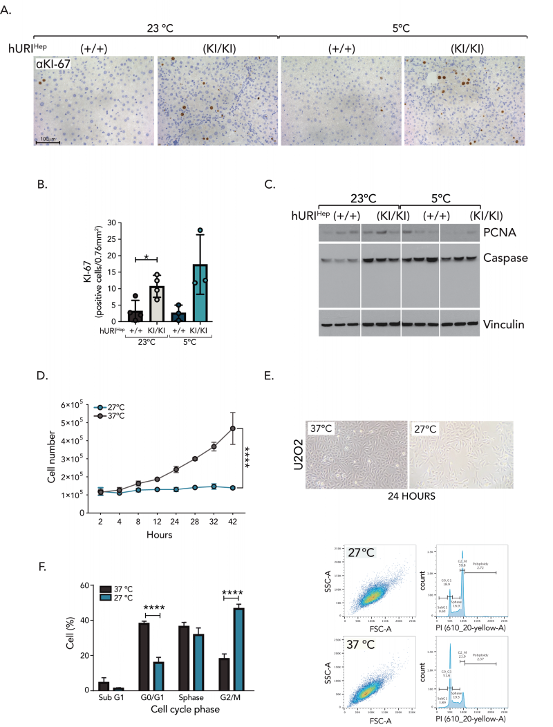
FIGURE 5: Cold exposure induces p53/p21-dependent G2/M cell cycle arrest. (A-B) (A) Representative pictures of KI67 IHC and (B) its quantification from liver sections of hURIHep mice exposed to RT or CT. (C) Western blot analysis of hURIHep mouse livers housed at RT or CT. (D) Cell number count of U2OS cells incubated for 24 hours at 27◦C or 37◦C [n=3]. (E) Representative image from bright field microscope of U2OS cells incubated for 24 hours at 27◦C or 37◦C. (F-G) Representative PI flow cytometry plots (F) and percentage of cell populations present at the different phases of the cell cycle (G). U2OS cells were incubated at 27◦C or 37◦C for 24 hours [n=3]. Statistical analysis was performed using Brown-Forsythe and Welch ANOVA (B), two-way ANOVA (D), and unpaired two-tailed Student’s t-test (F). Unless otherwise indicated, data are represented as mean ±s.e.m from n independent experiments; *P ≤ 0.05****P ≤ 0.001. Scale bars 100 µm (A).

