Back to article: Cold exposure reinstates NAD+ levels and attenuates hepatocellular carcinoma
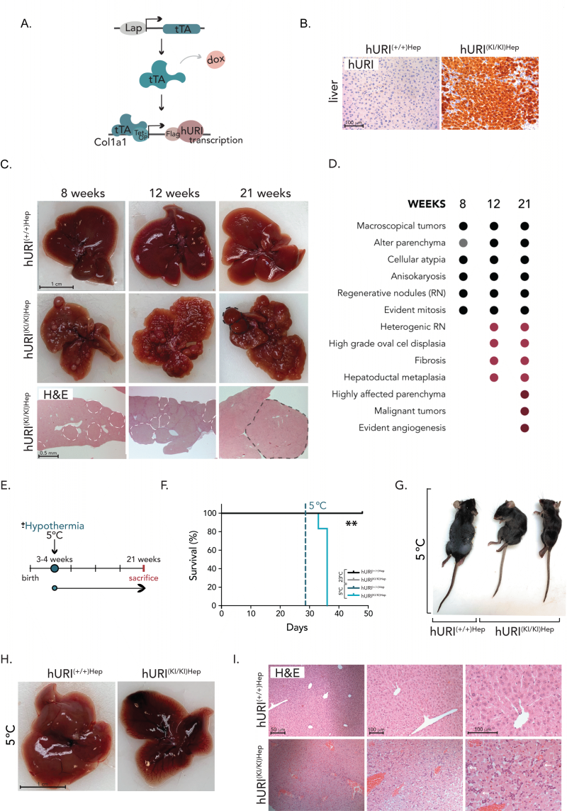
FIGURE 1: Hepatocytic hURI overexpression sensitizes mice to cold-induced stress. (A) Schematic representation of hURI-tetOFFHep (hURIHep) mouse model in which hURI expression is under the control of the hepatocyte-specific LAP promoter. (B) Representative pictures of immunohistochemistry (IHC) staining from liver sections of hURIHep mice highlighting hURI overexpression. (C) Representative images of livers from 8-,12-, and 21-week-old mice and representative pictures of hematoxylin and eosin (H&E) staining’s from hURI(+/+)Hep and hURI(KI/KI)Hep mice. (D) Histopathological characterization of 8-,12-, and 21-week-old hURI(KI/KI)Hepmice. (E) Scheme of hURIHep mice exposed to room temperature (RT) (23±1◦C) or cold temperature (CT) (5±1◦C) after weaning. (F) Survival curve of hURI(+/+)Hep [n=4], hURI(KI/KI)Hep [n=3] mice exposed to RT, and hURI(+/+)Hep [n=3] and hURI(KI/KI)Hep [n=6] mice exposed to CT. (G) Representative image of hURIHep mice exposed to RT and CT. (H) Representative images from hURIHep mice livers exposed to CT. (I) Representative pictures of H&E from liver sections of hURIHep mice exposed to CT. White arrows highlight oval cell hyperplasia. Statistical analysis was performed using the Kaplan-Meier curve (F). Unless otherwise indicated, data are represented as mean ± s.e.m from “n” independent experiments; **P ≤0.01. Scale bars, 50 µm, 100 µm (B, I), 0.5 mm (C), and 1 cm (C, H).