Back to article: Neuroglobin-enriched secretome provides neuroprotection against hydrogen peroxide and mitochondrial toxin-induced cellular stress
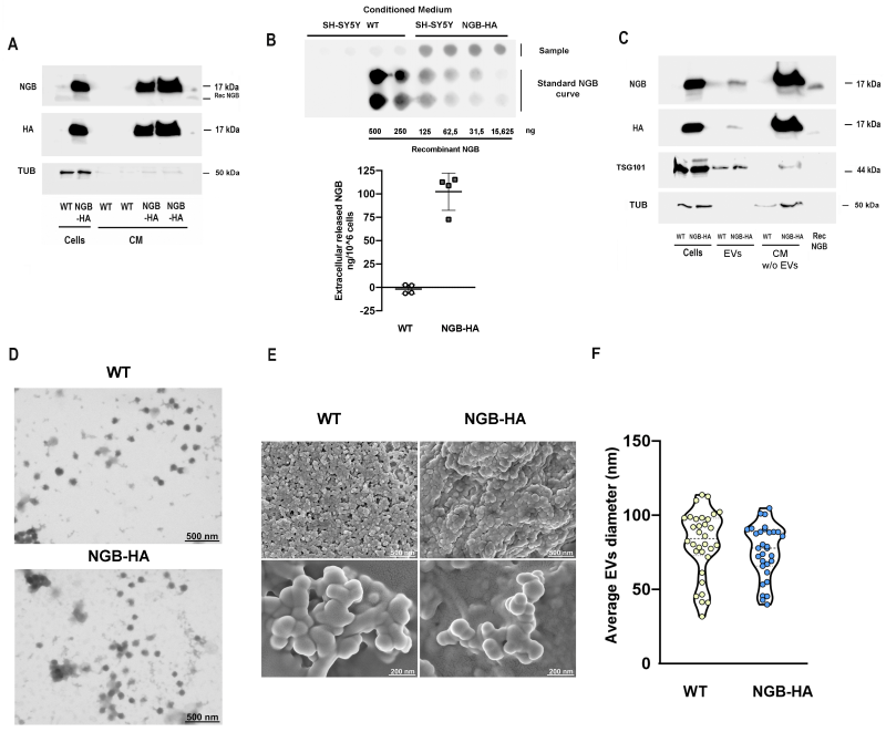
FIGURE 1: Effects of NGB overexpression on extracellular globin release. NGB and HA levels were measured by Western blot in conditioned medium (A) or in EVs and in conditioned medium deprived from EVs (CM w/o EVs) (C) obtained from SH-SY5Y wild type (WT) or overexpressing NGB-HA (NGB-HA) cells. Cell lysates were used as The levels of tubulin were evaluated on the same filter as cell lysate loading control or as marker of passively released protein in CM. The common exosomes marker TSG101 was used as loading control for EVs (C). Where indicated, recombinant NGB protein (2.5 ng) was used as a control. Blot images are representative of at least three independent experiments with similar results. Dot blot analysis (Top) and relative quantification of concentration (Bottom) of extracellular NGB in conditioned media obtained from WT and NGB-HA SH-SY5Y cells in quadruplicate (B). NGB recombinant concentration curve was used as dose reference for evaluating the concentration of extracellular NGB. Electron microscopy investigations confirmed the spheroidal morphology of EVs obtained from WT and NGB-HA overexpressing cells as can be observed both in representative STEM micrographs of EVs adsorbed on TEM grids (D) as well as in representative SEM micrographs of EVs mounted on silicon wafers (E). STEM images were used to analyze the diameter distribution of EVs obtained from WT and NGB-HA overexpressing cells (F).

