Back to article: Stress granules formation in HEI-OC1 auditory cells and in H4 human neuroglioma cells secondary to cisplatin exposure
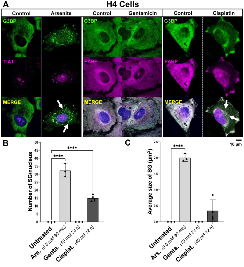
FIGURE 2: Influence of arsenite, gentamicin, and cisplatin on the induction of SG formation in H4 human neuroglioma cells. (A) Representative images of H4 human neuroglioma cells incubated with 0.5 mM arsenite for 30 min, 10 mM gentamicin for 24 h, or 40 µM cisplatin for 12 h, fixed and stained with a dual antibody combination targeting SG proteins anti-TIA1/PABP and anti-G3BP. The arrows point to SGs. The scale bar is 10 µm. (B) Bar graphs depicting variations in the quantified number of SG per cell, dependent upon treatment with arsenite, gentamicin or cisplatin, compared to control. (C) Bar graphs illustrating the average size of SG in µm2, influenced by incubation with arsenite, gentamicin or cisplatin, versus control. On the bar graphs, the x-axis represents the treatment condition, and the y-axis represents whether the number of SG per cell, or the size of SG in µm2 by the mean ± SD of n = 3 independent experiments for all treatment groups. Statistical analysis was performed using one-way ANOVA with Tukey’s multiple comparison test; with the adjusted p-value representing * = p < 0.05, ** = p < 0.01, *** = p < 0.001, **** = p < 0.0001.

