Back to article: The role of lipids in autophagy and its implication in neurodegeneration
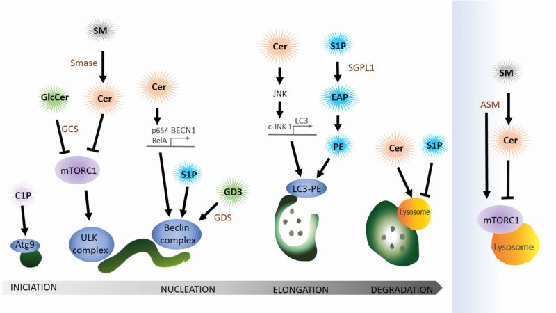
FIGURE 5: Bioactive sphingolipids, ceramide and sphingosine-1-phosphate (S1P) regulate autophagy especially in the initial steps. Lipids from the catabolic (blue), the sphingomyelin (grey) and hydrolytic (green) sphingolipid pathways function in the autophagy pathway. Sphingomyelin (SM) conversion to ceramide (Cer) by sphingomyelinase (SMase) affects mTORC1 activity on the lysosome and also on the phagophore. Cer can also induce autophagy by activating the transcription of ATG, BECN1 (Beclin) and LC3. Glycosphingolipids, glucosylceramide (GlcCer) and ganglioside (GD3) affect the autophagy pathway via mTORC1 or the Beclin complex respectively. LC3 conjugation to phosphatidylethanolamine (PE) is modulated by the catabolic sphingolipid pathway since sphingosine-1-phosphate (S1P) conversion to ethanolamine phosphate (EAP) that can be further converted to PE. During auto-lysosomal fusion Cer and SP1 have opposing effects (See text for details). Ceramide (Cer), sphingomyelin (SM), sphingomyelinase (SMase), acid sphingomyelinase (ASM), glucosylceramide (GlcCer), glucosylceramide synthase (GCS), ganglioside (GD3), sphingosine-1-phosphate (S1P), sphingosine-1-phosphate lyase 1 (SGPL1), ethanolamine phosphate (EAP), phosphatidylethanolamine (PE).

