Back to article: Mitochondria in cancer
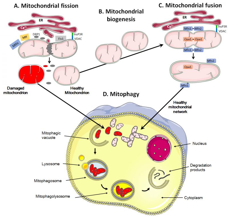
FIGURE 3: A high mitochondrial turnover rate is characteristic of many cancer cells. Mitochondrial quality control involves fission and mitophagy to eliminate defective mitochondria, whereas repopulation and functionalization depends on mitochondrial biogenesis and fusion. (A) During fission, the mitochondrion is marked and anchored by the endoplasmic reticulum (ER), notably through the binding of inositol 1,4,5-trisphosphate receptor (InsP3R) at the ER surface to voltage-dependent anion channel (VDAC) at the mitochondrial surface. This leads to the recruitment of dynamin-related protein 1 (DRP1), mitochondrial receptor protein 1 (Fis1), mitochondrial fission factor (Mff) and mitochondrial dynamic proteins (MIDs), allowing oligomerization and constriction to yield two daughter mitochondria. (B) During mitochondrial biogenesis, a mitochondrion self-replicates. (C) During fusion, mitofusins Mfn1 and Mfn2 are located on the outer mitochondrial membrane, allowing the exchange of calcium for signaling and creating antiparallel connections between two fusing mitochondria. Optic atrophy 1 (Opa1) together with Mnf1 participate in the fusion of the inner mitochondrial membrane. Fusion allows the formation of mitochondrial networks. (D) The mitophagic process consists in the engulfment of damaged mitochondria in a vacuole, called ‘mitophagic vacuole'. The subsequent fusion of the mitophagosome with lysosomes, forming a mitophagolysosome, allows the degradation of mitochondria in macromolecules that are delivered to the cytosol. Mitophagy can be non-selective or selective, using canonical and non-canonical pathways. It prevents the accumulation of damaged mitochondria that could harm or even kill the cell if apoptosis and/or the production of reactive oxygen species would derail.

