Back to article: Exocytotic fusion pore under stress
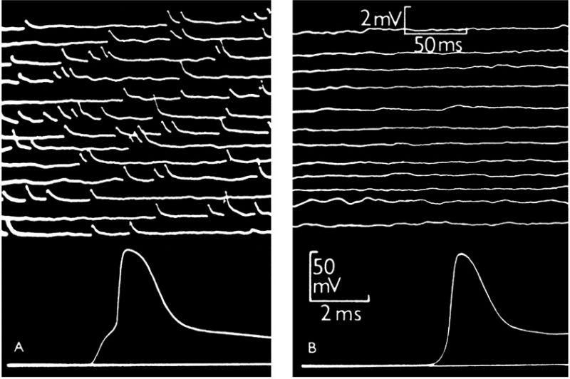
FIGURE 1: Intracellular recording from single muscle fibre of frog.(A) At the motor end-plate. The upper part shows spontaneous miniature end-plate potentials, which are localized at a junction and arise from sudden discharge by a motor nerve ending of packets of acetylcholine, each containing thousands of molecules. The lower part shows a single response to a nerve impulse, which was started by electric shock at the beginning of the trace; the first step of the response is large end-plate potential resulting from synchronous delivery of a few hundred packets of acetylcholine, this leading to full size action potential. (B) Traces recorded in the same muscle fibre, 2 mm away from the end-plate. The upper part shows much attenuated and barely recognizable residues of miniature end-plate potentials. The lower part shows propagated action potential, delayed by conduction over 2 mm distance and not preceded by the end-plate step. From [1]
1. Fatt PK, Katz B (1950). Some observations on biological noise. Nature 166(4223): 597-598. 10.1038/166597a0