Back to article: High mitochondrial calcium levels precede neuronal death in vivo in Alzheimer’s disease
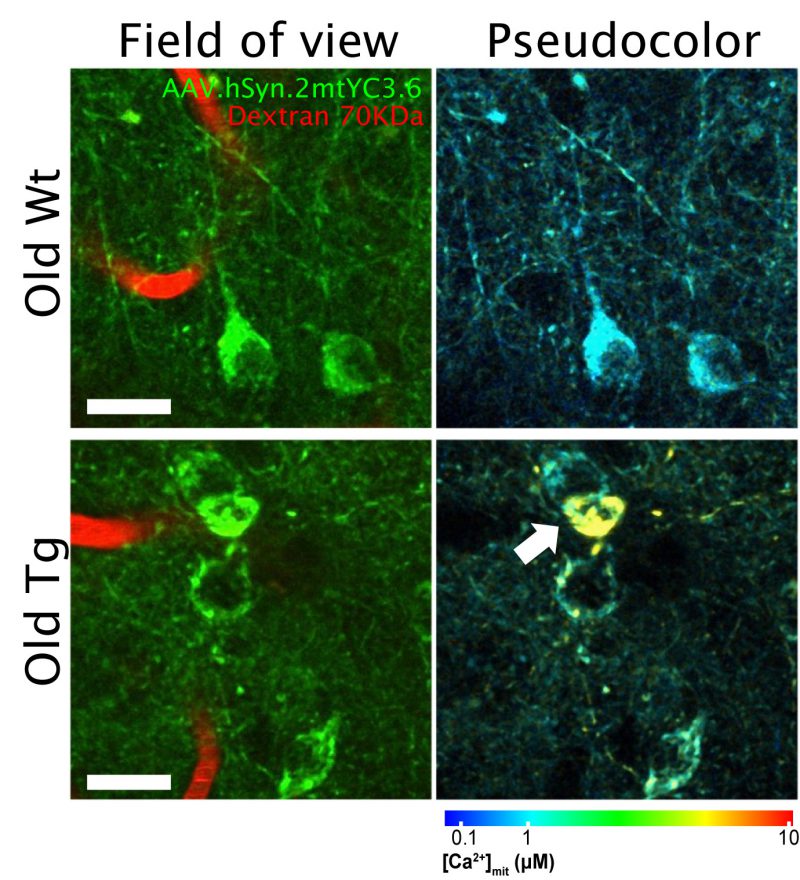
FIGURE 1: In vivo multiphoton microscopy images of mitochondrial Ca2+ in neurons in the Wt and APP/PS1 mice. Mice were injected with AAV.hSyn.2mtYC3.6 and a cranial window was implanted. Mitochondrial Ca2+ was later evaluated with multiphoton microscopy. Pictures are representative of old wild-type (Wt, top) and transgenic mice (APP/PS1 Tg, bottom). The field of view shows the AAV transduction in neuronal mitochondria (green) and the blood vessels labeled with fluorescent Dextran (red). Pseudocolor images represent the color coded mitochondrial Ca2+ concentrations for the corresponding field of view images. Arrow points to a neuron with mitochondrial Ca2+ overload. Scale bar represents 20 μm.