Back to article: LTX-315 sequentially promotes lymphocyte-independent and lymphocyte-dependent antitumor effects
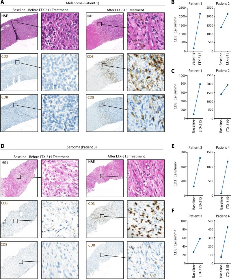
FIGURE 4: LTX-315 treatment causes CD8 T cell infiltration of melanoma and soft tissue sarcoma tumors in patients. (A) Representative hematoxylin and eosin (H&E) staining and immunohistochemistry for CD3+ and CD8+ cells (brown) on melanoma patient biopsy sections before and after LTX-315 treatment. (B) Quantification of CD3+ cells in melanoma biopsies, based on immunohistochemistry staining shown in (A). (C) Same analysis as shown in (B) for quantification of CD8+ cells. (D) Representative H&E staining and immunohistochemistry for CD3+ and CD8+ cells (brown) on patient biopsy sections of sarcomas before and after LTX-315 treatment. (E) Quantification of CD3+ cells in sarcoma biopsies, based on immunohistochemistry staining shown in (D). (F) Same analysis as shown in (E) for quantification of CD8+ cells.