Back to article: Gold Nanoparticles sensitize pancreatic cancer cells to gemcitabine
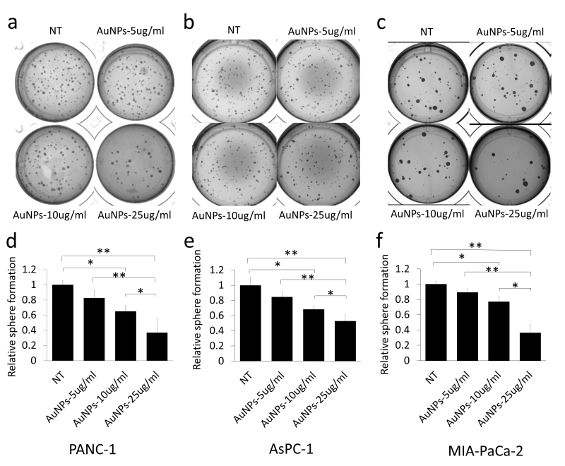
FIGURE 3: Dose dependent inhibition of 3D sphere formation in AuNPs treated pancreatic cancer cell lines. PANC-1 (a) (600 cells/well), AsPC-1 (b) (600 cells/well) and MIA-PaCa-2 (c) (400 cells/well) were suspended in complete medium and then mixed with equal volumes of matrigel, the cell mixture was added on the top of the lower layer of solid matrix (complete medium: matrigel=1:1) in 24 wells plate. On the top the mixture, 1 ml of complete medium with different concentrations of AuNPs (5 μg/ml, 10 μg/ml or 25 μg/ml) was added. The sphere numbers were counted 20-40 days after seeding. (a-c) The spheres formed with different pancreatic cancer cells with different concentration of AuNPs treated. (d-f) Relative sphere numbers corresponding to a-c. Independent experiments were repeated for at least three times and each time, at least triplicate wells were used. Values are means ±SD and statistical analysis were performed using one-way ANOVA. *P≤0.05, **P≤0.01.

