Table of contents
Volume 3, Issue 12, pp. 361 - 387, December 2019
Cover: This month in
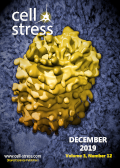
Cell Stress: Golgi and ER stress in microcephaly. Image shows 3D reconstruction of a Golgi body (yellow), based on crossbeam FIB-SEM ion milling raw data. Courtesy of Louise Hughes, Oxford Brookes University, UK, licensed under a
CC BY 2.0 license. Modified by
Cell Stress. The cover is published under the
CC BY 4.0 license.
Enlarge issue cover
Endoplasmic reticulum and Golgi stress in microcephaly
Sandrine Passemard, Franck Perez, Pierre Gressens and Vincent El Ghouzzi
Reviews |
page 369-384 | 10.15698/cst2019.12.206 | Full text | PDF |
Abstract
Microcephaly is a neurodevelopmental condition characterized by a small brain size associated with intellectual deficiency in most cases and is one of the most frequent clinical sign encountered in neurodevelopmental disorders. It can result from a wide range of environmental insults occurring during pregnancy or postnatally, as well as from various genetic causes and represents a highly heterogeneous condition. However, several lines of evidence highlight a compromised mode of division of the cortical precursor cells during neurogenesis, affecting neural commitment or survival as one of the common mechanisms leading to a limited production of neurons and associated with the most severe forms of congenital microcephaly. In this context, the emergence of the endoplasmic reticulum (ER) and the Golgi apparatus as key guardians of cellular homeostasis, especially through the regulation of proteostasis, has raised the hypothesis that pathological ER and/or Golgi stress could contribute significantly to cortical impairments eliciting microcephaly. In this review, we discuss recent findings implicating ER and Golgi stress responses in early brain development and provide an overview of microcephaly-associated genes involved in these pathways.
Metabolism remodeling in pancreatic ductal adenocarcinoma
Jin-Tao Li, Yi-Ping Wang, Miao Yin and Qun-Ying Lei
Reviews |
page 361-368 | 10.15698/cst2019.12.205 | Full text | PDF |
Abstract
Pancreatic ductal adenocarcinoma (PDAC) is predicted to become the second leading cause of death of patients with malignant cancers by 2030. Current options of PDAC treatment are limited and the five-year survival rate is less than 8%, leading to an urgent need to explore innovatively therapeutic strategies. PDAC cells exhibit extensively reprogrammed metabolism to meet their energetic and biomass demands under extremely harsh conditions. The metabolic changes are closely linked to signaling triggered by activation of oncogenes like KRAS as well as inactivation of tumor suppressors. Furthermore, tumor microenvironmental factors including extensive desmoplastic stroma reaction result in series of metabolism remodeling to facilitate PDAC development. In this review, we focus on the dysregulation of metabolism in PDAC and its surrounding microenvironment to explore potential metabolic targets in PDAC therapy.
Stress granules regulate paraspeckles: RNP granule continuum at work
Haiyan An and Tatyana A. Shelkovnikova
Microreviews |
page 385-387 | 10.15698/cst2019.12.207 | Full text | PDF |
Abstract
Eukaryotic cells contain several types of RNA-protein membraneless macro-complexes – ribonucleoprotein (RNP) granules that form by liquid-liquid phase separation. These structures represent biochemical microreactors for a variety of cellular processes and also act as highly accurate sensors of changes in the cellular environment. RNP granules share multiple protein components, however, the connection between spatially separated granules remains surprisingly understudied. Paraspeckles are constitutive nuclear RNP granules whose numbers significantly increase in stressed cells. Our recent work using affinity-purified paraspeckles revealed that another type of RNP granule, cytoplasmic stress granule (SG), acts as an important regulator of stress-induced paraspeckle assembly. Our study demonstrates that despite their residency in different cellular compartments, the two RNP granules are closely connected. This study suggests that nuclear and cytoplasmic RNP granules are integral parts of the intracellular “RNP granule continuum” and that rapid exchange of protein components within this continuum is important for the temporal control of cellular stress responses. It also suggests that cells can tolerate and efficiently handle a certain level of phase separation, which is reflected in the existence of “bursts”, or “waves”, of RNP granule formation. Our study triggers a number of important questions related to the mechanisms controlling the flow of RNP granule components within the continuum and to the possibility of targeting these mechanisms in human disease.


