Table of contents
Volume 1, Issue 3, pp. 110 - 140, December 2017
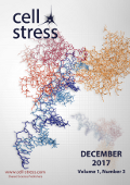
Cover: This month in
Cell Stress: DNA replication stress. Structure of human Poly(ADP-ribose)-Polymerase (PARP-1) bound to a DNA (blue) double strand break. PDB entry
4DQY by M.F. Langelier, J.L. Planck, S. Roy, and J.M. Pascal modified by
Cell Stress. The cover is published under the
CC BY 4.0 license.
Enlarge issue cover
Metastatic-initiating cells and lipid metabolism
Salvador Aznar Benitah
News and thoughts |
page 110-114 | 10.15698/cst2017.12.113 | Full text | PDF |
Abstract
The identity of the cells responsible for initiating and promoting metastasis has been historically elusive. Consequently, this has hampered our ability to develop specific anti-metastatic treatments, resulting in the majority of metastatic cancers remaining clinically untreatable. Furthermore, advances in genome sequencing indicate that the acquisition of metastatic competency does not seem to involve the accumulation of de novo mutations, making it difficult to understand why some tumours become metastatic while others do not. We have recently identified metastatic-initiating cells, and described how they specifically rely on fatty acid uptake and lipid metabolism to promote metastasis. This intriguing finding indicates that external influences, such as those derived from our diet, exert a strong influence on tumour progression, and that such dietary factors could be therapeutically modulated if understood. In this News and Thoughts, I will comment on recent findings regarding how and why lipid metabolism modulates the behaviour of metastatic cells, and how this knowledge can be harnessed to devise new and specific anti-metastatic therapies.
A game of substrates: replication fork remodeling and its roles in genome stability and chemo-resistance
Julia Sidorova
Reviews |
page 115-133 | 10.15698/cst2017.12.114 | Full text | PDF |
Abstract
During the hours that human cells spend in the DNA synthesis (S) phase of the cell cycle, they may encounter adversities such as DNA damage or shortage of nucleotides. Under these stresses, replication forks in DNA may experience slowing, stalling, and breakage. Fork remodeling mechanisms, which stabilize slow or stalled replication forks and ensure their ability to continue or resume replication, protect cells from genomic instability and carcinogenesis. Fork remodeling includes DNA strand exchanges that result in annealing of newly synthesized strands (fork reversal), controlled DNA resection, and cleavage of DNA strands. Defects in major tumor suppressor genes BRCA1 and BRCA2, and a subset of the Fanconi Anemia genes have been shown to result in deregulation in fork remodeling, and most prominently, loss of kilobases of nascent DNA from stalled replication forks. This phenomenon has recently gained spotlight as a potential marker and mediator of chemo-sensitivity in cancer cells and, conversely, its suppression – as a hallmark of acquired chemo-resistance. Moreover, nascent strand degradation at forks is now known to also trigger innate immune response to self-DNA. An increasingly sophisticated molecular description of these events now points at a combination of unbalanced fork reversal and end-resection as a root cause, yet also reveals the multi-layered complexity and heterogeneity of the underlying processes in normal and cancer cells.
New insight into the role of autophagy in tumorigenesis
Yongjun Tian, Linya Wang and Jing-hsiung James Ou
Microreviews |
page 136-138 | 10.15698/cst2017.12.116 | Full text | PDF |
Abstract
Autophagy plays an important role in maintaining cellular homeostasis. Its dysfunction can cause many diseases, including neurodegenerative diseases, metabolic diseases and cancer. The role of autophagy in carcinogenesis is complex, as it was shown to have pro-tumorigenic functions in some reports, but anti-tumorigenic functions in others. By using mice with hepatocyte-specific knockout of Atg5, a gene essential for autophagy, we had previously demonstrated that impairing autophagy in hepatocytes would induce oxidative stress and DNA damage, followed by the initiation of hepatocarcinogenesis. Interestingly, these mice developed only benign tumors with no hepatocellular carcinoma (HCC), even after they were treated with the carcinogen diethylnitrosamine (DEN), which induced HCC in wild-type mice. Our recent studies indicated that the inability of mice to develop HCC when autophagy was impaired was at least partially due to the activation of the tumor suppressor TP53, which suppressed the expression of NANOG, a transcription factor critical for the self-renewal and the maintenance of cancer stem cells (CSCs).
Protein quality control meets transcriptome remodeling under stress
Veena Mathew and Peter C. Stirling
Microreviews |
page 134-135 | 10.15698/cst2017.12.115 | Full text | PDF |
Abstract
To tolerate and recover from genotoxic stress cells must coordinate a range of stress response activities including cell cycle arrest, DNA repair, and remodeling of the transcriptome and proteome. The suppression of ribosome production is a key feature of many stress responses in yeast, and much is known about the dynamics of this process at the transcriptional level. In our recent study, (J Cell Biol doi: 10.1083/ jcb.201612018) we focus on the stress related dynamic behaviour of a splicing factor called Hsh155, which is a core component of the SF3B subcomplex of the U2 small nuclear ribonucleoprotein complex, homologous to human SF3B1. The disassembly from its complex and sequestration of Hsh155 into nuclear protein aggregates contributes to suppressing ribosome production post-transcriptionally by promoting intron retention in ribosomal protein gene transcripts. The relocalization of Hsh155 is facilitated by TORC1-driven transcriptional changes and molecular chaperones that recognize disassembled Hsh155, eventually aiding in efficient recovery from stress.
Reciprocal regulatory circuits coordinating EMT plasticity
Maren Diepenbruck and Gerhard Christofori
Microreviews |
page 139-140 | 10.15698/cst2017.12.117 | Full text | PDF |
Abstract
Epithelial to mesenchymal transition (EMT) as well as its reversal process, mesenchymal to epithelial transition (MET), are essential and well-controlled cellular processes during embryonic development. Tightly controlled regulatory mechanisms guide an EMT/MET plasticity and enable cells to switch forth and back between different cell morphologies and functional capabilities to endow the necessity of tissue plasticity. However, aberrant and uncontrolled activation of these processes during malignant tumor progression promotes primary tumor cell invasion, cancer cell dissemination and metastatic outgrowth. In a recent study (Nat Commun; doi: 10.1038/s41467-017-01197-w), we have reported on the post-transcriptional control of normal and cancer-associated EMT by miRNAs and identified a novel, critical double-negative feedback regulation of the thus far unknown miRNA miR1199 and the key EMT transcription factor Zeb1.


