Table of contents
Volume 2, Issue 8, pp. 184 - 218, August 2018
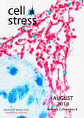
Cover: This month in
Cell Stress: Mitochondrial dysfunction and cellular stress. Image depicts mitochondrial network (red) surrounding the nucleus (blue). Credit: NICHD/U. Manor, licensed under a
CC BY 2.0 license. Image modified by
Cell Stress. The cover is published under the
CC BY 4.0 license.
Enlarge issue cover
Mitochondrial dysfunction and its role in tissue-specific cellular stress
David Pacheu-Grau, Robert Rucktäschel and Markus Deckers
Reviews |
page 184-199 | 10.15698/cst2018.07.147 | Full text | PDF |
Abstract
Mitochondrial bioenergetics require the coordination of two different and independent genomes. Mutations in either genome will affect mitochondrial functionality and produce different sources of cellular stress. Depending on the kind of defect and stress, different tissues and organs will be affected, leading to diverse pathological conditions. There is no curative therapy for mitochondrial diseases, nevertheless, there are strategies described that fight the various stress forms caused by the malfunctioning organelles. Here, we will revise the main kinds of stress generated by mutations in mitochondrial genes and outline several ways of fighting this stress.
T lymphocytes against solid malignancies: winning ways to defeat tumours
Ignazio Caruana, Luca Simula, Franco Locatelli and Silvia Campello
Reviews |
page 200-212 | 10.15698/cst2018.07.148 | Full text | PDF |
Abstract
In the last decades, a novel field has emerged in the cure of cancer, by boosting the ability of the patient’s immune system to recognize and kill tumour cells. Although excellent and encouraging results, exploiting the effect of genetically modified T cells, have been obtained, it is now evident that tumour malignancies can evolve several mechanisms to escape such immune responses, thus continuing their growth in the body. These mechanisms are in part due to tumour cell metabolic or genetic alterations, which can render the target invisible to the immune system or can favour the generation of an extracellular milieu preventing immune cell infiltration or cytotoxicity. Such mechanisms may also involve the accumulation inside the tumour microenvironment of different immune-suppressive cell types, which further down-regulate the activity of cytotoxic immune cells either directly by interacting with them or indirectly by releasing suppressive molecules. In this review, we will first focus on describing several mechanisms by which tumour cells may dampen or abrogate the immune response inside the tumour microenvironment and, second, on current strategies that are adopted to cope with and possibly overcome such alterations, thus ameliorating the efficacy of the current-in-use anti-cancer immuno-therapies.
GFI1’s role in DNA repair suggests implications for tumour cell response to treatment
Charles Vadnais and Tarik Möröy
Microreviews |
page 213-215 | 10.15698/cst2018.07.149 | Full text | PDF |
Abstract
Despite recent advances in cancer treatment through personalized and precision medicine and new avenues such as immunotherapy and chimeric antibodies, the induction of DNA damage either through irradiation or specific compounds remains the primary approach to kill tumour cells. Improvements in our understanding of how tumour cells respond to DNA damage, and especially how this response differs from that of normal cells, are crucial to the development of better and more efficient therapies. We have recently shown that the activity of the oncogenic transcription factor GFI1, which is required for the development and maintenance of T and B cell leukemia, increases the ability of tumour cells to repair their DNA following damage (Vadnais et al. Nat Commun 9(1):1418). GFI1 accomplishes this by regulating the post-translational modifications (PTM) of key DNA repair proteins, including MRE11 and 53BP1, by the methyltransferase PRMT1. Here, GFI1 acts as an accessory protein required for the interaction between the enzyme and its substrates. This has implications for the treatment response of tumour cells overexpressing GFI1, which includes T cell leukemia, neuroendocrine lung carcinomas and aggressive subtypes of medulloblastoma, and suggests that targeting GFI1’s activity and with this its capacity to aid DNA repair may open avenues for new therapeutic approaches.
Irreversible impact of chronic hepatitis C virus infection on human natural killer cell diversity
Benedikt Strunz, Julia Hengst, Heiner Wedemeyer and Niklas K. Björkström
Microreviews |
page 216-218 | 10.15698/cst2018.07.150 | Full text | PDF |
Abstract
Diversity is crucial for the immune system to efficiently combat infections. Natural killer (NK) cells are innate cytotoxic lymphocytes that contribute to the control of viral infections. NK cells were for long thought to be a homogeneous population of cells. However, recent work has instead revealed NK cells to represent a highly diverse population of immune cells where a vast number of subpopulations with distinct characteristics exist across tissues. However, the degree to which a chronic viral infection affects NK cell diversity remains elusive. Hepatitis C virus (HCV) is effective in establishing chronic infection in humans. During the last years, new direct-acting antiviral drugs (DAA) have revolutionized treatment of chronic hepatitis C, enabling rapid cure in the majority of patients. This allows us to study the influence of a chronic viral infection and its subsequent elimination on the NK cell compartment with a focus on NK cell diversity. In our recent study (Nat Commun, 9:2275), we show that chronic HCV infection irreversibly impacts human NK cell repertoire diversity.


