Back to article: The role of hydrophobic matching on transmembrane helix packing in cells
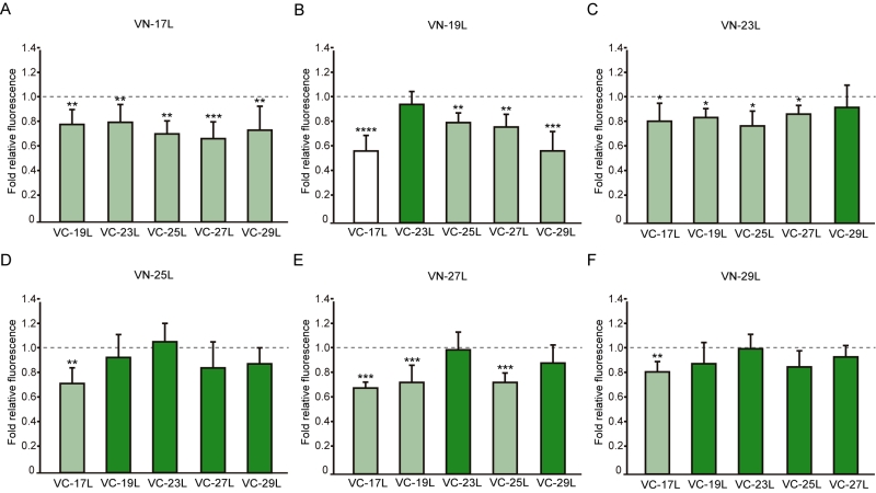
FIGURE 7: Differences in hetero-dimerization in eukaryotic membranes. Mean relative fluorescence of chimera hetero-oligomerization. For any given combination (eg.VN-X/VC-Y) the corresponding homo-oligomerization values (VN-X/VC-X and VN-Y/VC-Y) were used as a reference set to obtain fold change and significance (q-values,*<0.05, **<0.01, ***< 0.001, **** <0.0001). (A) VN-17L/VC-X, (B) VN-19L/VC-X, (C) VN-23L/VC-X, (D) VN-25L/VC-X, (E) VN-27L/VC-X, (F) VN-29L/VC-X. Error bars indicate the standard deviation obtained from at least 4 independent replicates. A color intensity code was used to highlight the dimerization intensity. Dark green (fluorescence values equivalent to the appropriated control), light green (fluorescence values significantly higher than those obtained with the H2 control but significantly lower than those observed with the corresponding homo-oligomer controls), and white (values equivalent to those obtained with the H2 control).

