Back to article: Dram1 regulates DNA damage-induced alternative autophagy
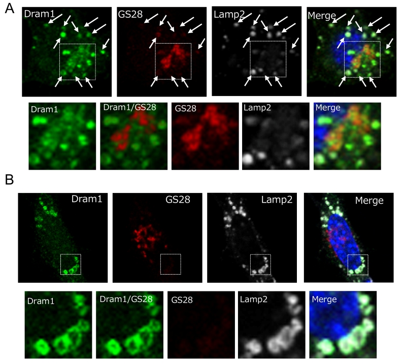
FIGURE 4: Intracellular localization of Dram1-Flag in Atg5 KO MEFs. Atg5 KO MEFs were transfected with dram1-flag for 24 h (A) and 48 h (B). The cells were then examined for immunofluorescence of GS28 (a pan-Golgi marker), Lamp2 (a lysosome marker), and Flag (Dram1). Nuclei were counterstained with DAPI. In (A), some Dram1 particles were merged with GS28 signals (dashed squares). Magnified images of the area within the dashed squares are shown in the bottom panels. Ar-rows indicate areas of Dram1 and Lamp2 colocalization. In (B), none of the Dram1 particles were merged with GS28 signals (dashed squares). Magnified images of the area within the dashed squares are shown in the bottom panels.

