Back to article: The role of hydrophobic matching on transmembrane helix packing in cells
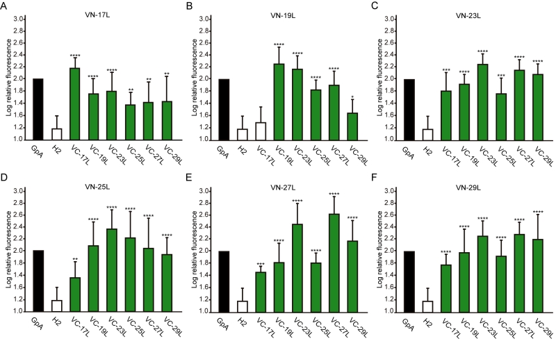
FIGURE 6: Hetero-dimerization in eukaryotic membranes. Mean relative fluorescence of chimera hetero-oligomerization in HEK293T cells of all different combinations: (A) VN-17L/VC-X, (B) VN-19L/VC-X, (C) VN-23L/VC-X, (D) VN-25L/VC-X, (E) VN-27L/VC-X, (F) VN-29L/VC-X. Error bars indicate standard deviation obtained from at least 4 independent replicates. GpA homo-dimer (black bars) was used as positive control and normalization value while Lep H2 homo-oligomer was used as a negative control (white bars). For the experimental samples, a color intensity code was used to highlight dimerization (green, fluorescence values significantly higher than those obtained with the H2 control) and non-dimerization (white, values equivalents to those obtained with the negative control). Additionally asterisks were included to indicate the level of significance (**<0.01, ***< 0.001, **** <0.0001).

